17 Oct 2019
Abstract
African swine fever virus (ASFV) is a giant and complex DNA virus that causes a highly contagious and often lethal swine disease without vaccine available. Using an optimized image reconstruction strategy, we solved the ASFV capsid structure up to 4.1-angstroms, which is built from 17,280 proteins, including one major (p72) and four minor capsid proteins (M1249L, p17, p49 and H240R), organized into pentasymmetrons and trisymmetrons. The atomic structure of the p72 informs putative conformational epitopes, distinguishing ASFV from other nucleocytoplasmic large DNA viruses (NCLDV). The minor capsid proteins form a complicated network below the outer capsid shell, stabilizing the capsid by holding adjacent capsomers together. Acting as core organizers, 100-nm long M1249L proteins run along each edge of trisymmetrons bridging two neighboring pentasymmtrons and form extensive intermolecular networks with other capsid proteins, driving the formation of the capsid framework. These structural details unveil the basis of capsid stability and assembly, opening up new avenues for ASF vaccine development.
[Image]
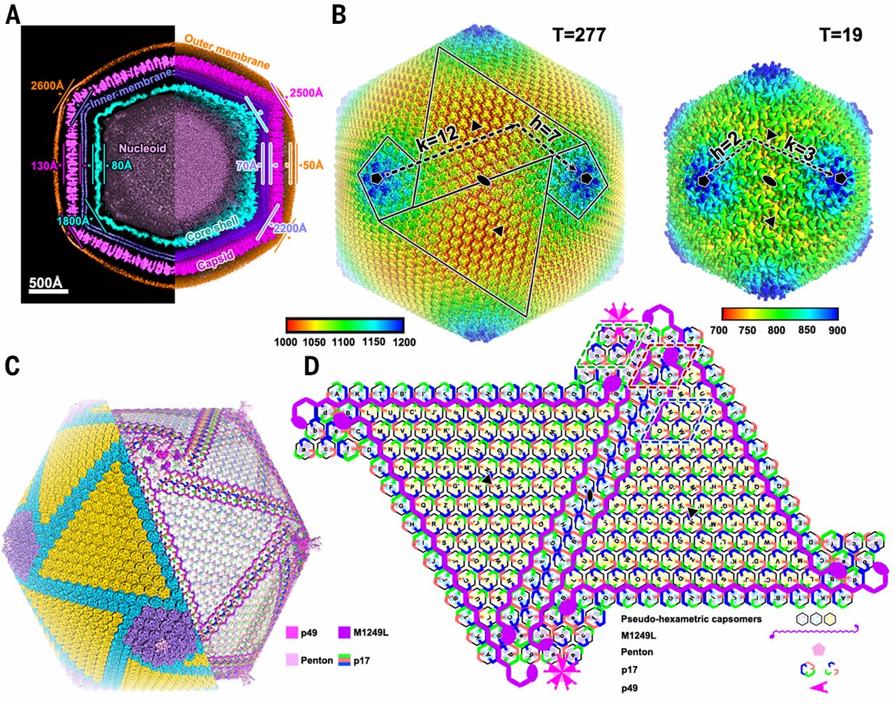
Fig. 1 Architecture of the ASFV virion.
(A) The central slice (left) and cross-section (right) of the icosahedral ASFV virion structure. The outer membrane, capsid, inner membrane, core shell, and nucleoid are colored in orange, magenta, deep-blue, cyan, and grey, respectively. The radius and thickness of each layer are labeled.
(B) Radially colored representations of the ASFV capsid and core shell. The T number including the h and k vectors are indicated.
(C) Cryo-EM reconstruction of the ASFV capsid. Left half: trisymmetron and pentasymmetry organization, the trisymmetrons, pentasymmetrons and zippers (the boundaries of two neighboring trisymmetrons) are colored in yellow, light-purple and cyan respectively. Right half: density of the minor capsid proteins, including the penton proteins after removing the outer capsid shell, each minor capsid protein is shown in a different color as indicated on the right. Boxes in green, red and blue show the locations with the representative capsomer assembly patterns, that will be discussed in Fig. 2D.
(D) Diagrammatic organization of the minor capsid proteins and capsomers viewed from inside capsid. The pseudo-hexameric capsomers are outlined. The icosahedral 3-fold and 2-fold axes are shown as solid black triangles and ovals, respectively. Different minor capsid proteins including the penton proteins are shown as different shapes with different colors, as indicated. The pseudo-hexameric capsomers are labeled A, B, C, ... in the trisymmetrons and a, b, c, ... in the pentasymmetrons.